


Introduction To Skin Lesion Identification And Dermatoscopy
Introduction To Skin Lesion Identification And Dermatoscopy
-
Chapter 1 : Pre Course Knowledge Evaluation
-
Chapter 2 : What To Expect
-
Chapter 3 : Introduction To Skin AssessmentLecture 1: Before We BeginPrivateLecture 2: Summary Of Skin Assessment ProtocolsPrivateLecture 3: Scanning TechnologyPrivateLecture 4: Scanning Technology: Wood's LampPrivateLecture 5: Benefits Of Thorough Skin Assessment And AnalysisPrivateLecture 6: Limitations of the Skin AssessmentPrivateLecture 7: How do we identify skin lesions?PrivateLecture 8: Identification of Skin LesionsPrivate
-
Chapter 4 : Introducing DermatoscopyLecture 1: Introducing DermatoscopyPrivateLecture 2: Dermatoscopy: The Gold StandardPrivateLecture 3: Art Of DermatoscopyPrivateLecture 4: The Importance Of Dermatoscopy in Clinical PracticePrivateLecture 5: Introducing the DermatoscopePrivateLecture 6: Polarised Immersion TechniquesPrivate
-
Chapter 5 : Benign Skin Lesion IdentificationLecture 1: Review Of The Most Common Benign Melanocytic And Non-Melanocytic LesionsPrivateLecture 2: Melanocytic LesionsPrivateLecture 3: Melanocytic Lesions : NaeviPrivateLecture 4: Non-Melanocytic LesionsPrivateLecture 5: Melanotic MaculesPrivateLecture 6: Ephelides (Freckles)PrivateLecture 7: Vascular LesionsPrivateLecture 8: Common Benign Lesions and Conditions On The Face, Neck, Scalp & DécolletagePrivateLecture 9: Examples Of Conditions Found On The Head And NeckPrivateLecture 10: Examples Of Conditions Found On The Head And NeckPrivateLecture 11: Examples Of Conditions Found On The Head And FacePrivateLecture 12: Commonly Occurring Benign Lesions And Conditions On The Torso & Upper LimbsPrivateLecture 13: Examples Of Lesions Found On The Head, Trunk and Upper LimbsPrivateLecture 14: Examples Of Lesions Found On The Trunk And Upper LimbsPrivateLecture 15: Examples Of Conditions Found On The Trunk And Upper LimbsPrivateLecture 16: Examples Of Conditions Found On The Trunk And Upper LimbsPrivateLecture 17: Commonly Found Benign Lesions And Conditions On The Buttocks, Legs And FeetPrivateLecture 18: Lesions Found On The Legs, Buttocks And FeetPrivateLecture 19: Lesions Found On The Legs, Buttocks And Feet (2)PrivateLecture 20: Variations Of Seborrheic KeratosisPrivateLecture 21: Progression of Seborrhoeic KeratosisPrivateLecture 22: Dermatoscopic Images of Benign Melanocytic LesionsPrivateLecture 23: Dermatoscopic Images Of Benign Non-Melanocytic LesionsPrivate
-
Chapter 6 : Malignant Skin Lesion IdentificationLecture 1: IntroductionPrivateLecture 2: The ABCDE AlgorithmPrivateLecture 3: Pattern Analysis: When The ABCDE Rule Isn’t EnoughPrivateLecture 4: Dermatoscopic Criteria DevelopmentPrivateLecture 5: Pattern Analysis MethodsPrivateLecture 6: The “Blink” TestPrivateLecture 7: The Three-Step ChecklistPrivateLecture 8: STOLZ ABCD RulePrivateLecture 9: The Menzies’ MethodPrivateLecture 10: Establishing Confirmation Of Lesions – Chaos And Clues AlgorithmPrivateLecture 11: Skin Cancer – Non-Melanoma Skin Cancer (NMSC)PrivateLecture 12: Basal Cell Carcinoma (BCC)PrivateLecture 13: Basal Cell Carcinoma (BCC) IdentificationPrivateLecture 14: Pigmented Basal Cell Carcinoma (PBCC)PrivateLecture 15: Basal Cell Carcinoma (BCC) - Dermatoscopic FeaturesPrivateLecture 16: Basal Cell Carcinoma (BCC) - Dermastoscopic FeaturesPrivateLecture 17: Nodular Basal Cell Carcinoma (BCC)PrivateLecture 18: Actinic Keratosis (Solar Keratosis)PrivateLecture 19: Actinic KeratosisPrivateLecture 20: Features Of Actinic KeratosisPrivateLecture 21: Intraepidermal Carcinoma (IEC)/ SCC In SITU/Bowens DiseasePrivateLecture 22: Squamous Cell CarcinomaPrivateLecture 23: Squamous Cell Carcinoma – Well DifferentiatedPrivateLecture 24: Squamous Cell Carcinoma of the LipPrivateLecture 25: Squamous Cell Carcinoma - Dermatoscopic FeaturesPrivateLecture 26: Malignant MelanomaPrivateLecture 27: Malignant MelanomaPrivateLecture 28: Malignant Melanoma (3)PrivateLecture 29: Lentigo Maligna MelanomaPrivateLecture 30: Subungual Lentiginous MelanomaPrivateLecture 31: Subungual MelanomaPrivateLecture 32: Superficial Spreading Melanoma – AmelanoticPrivateLecture 33: Amelanotic MelanomaPrivateLecture 34: Acral Lentiginous Patterns – Lesions Of The Volar Skin (Soles Of The Feet/PalmsPrivateLecture 35: Acral Lentiginous MelanomaPrivateLecture 36: SummaryPrivateLecture 37: A Summary of Skin Lesion DermatoscopyPrivate
-
Chapter 7 : Self Assessment
-
Chapter 8 : The Referral Process
-
Chapter 9 : Conclusion
-
Chapter 10 : Reference List
-
Chapter 11 : Final Assessment
-
Chapter 12 : Participants Feedback Form
Course Description
CPD: 2 Hours
Location: Online Only
Certificate: Certificate of Completion
Prerequisite: Fundamentals of Skin Science (Recommended)
Suitable For: All Professions
Price: $88 Lifetime Access
Introduction To Skin Lesion Identification And Dermatoscopy has been developed for cosmetic practitioners to have an opportunity to learn to identify simple and complex skin lesions, including skin cancers and the use of the dermatoscope as a diagnostic tool.
It can be very challenging when one is presented with a mole on the patient’s treatment area and not knowing whether to proceed with the treatment or ask the patient to seek further advise with their doctor before proceeding with further treatment. This module does not aim to replace the skin doctor nor their experience but rather give you the basic understanding of what to look out for and what certain signs mean, including those seen under the dermatoscope. Dermatoscopy is essential skill in cosmetic dermatology and each cosmetic practitioner should be well versed in it.
We recommend that you complete Fundamentals of Skin Science before attempting this module as it will set a good foundation in skin physiology which is important to better understand how skin lesions are identified.
Want to accelerate your learning? Save 15% when you purchase Introduction To Skin Lesion Identification And Dermatoscopy in a bundle:
Learning Objectives
Upon completion of this module, you should be able to:
- Understand the importance and use of dermatoscopy in a skin therapy framework
- Be able to identify common benign lesions based on simple algorithms
- Understand the difference between a benign lesion and a malignant lesion
- Have an understanding of the ABCDE algorithm and how to apply it
- Have an introduction into the Chaos and Clues algorithm
- Be able to establish cross-referral protocols for patient safety and clinical best practice
Team
The Aesthetics and Skin Institute (ASI) is a global training organisation based in Australia. Our mission is to empower your success in aesthetics, whether you are a doctor, dentist, nurse or skin therapist. We offer both online courses, hands-on training and certificate programs to help you become a confident practitioner in cosmetic injectables, threads, lasers, skin therapy and much more.
Led by Dr Ben Chan, our team of educators are successful industry leaders who are passionate about teaching and pushing the boundaries of aesthetics medicine.
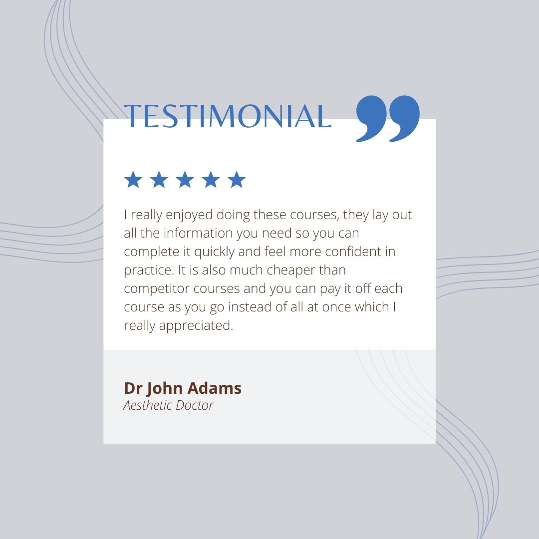
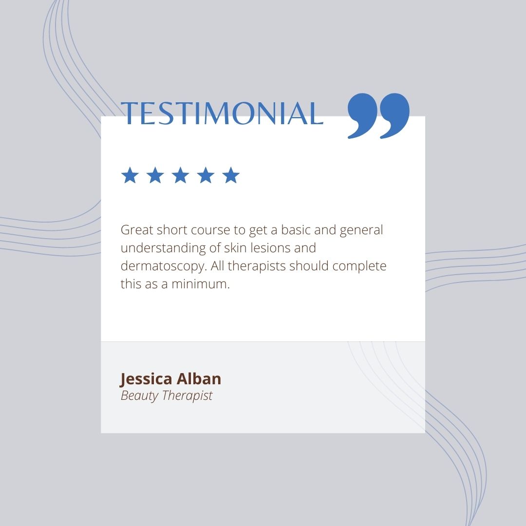
Testimonials
 |  |
Login to see discussions
Highlights
- Understand the benefits of dermatoscopy in a skin therapy framework
- Be able to identify common benign lesions based on simple algorithms
- Understand the difference between a benign lesion and a malignant lesion
- Have an understanding of the ABCDE algorithm and how to apply this
- Know what to do if a lesion does not fit the ABCDE algorithm
Related Courses
.jpeg)